
Overview- Echocardiogram
Echocardiogram
An echocardiogram uses sound waves to produce images of your heart. This common test allows your doctor to see your heart beating and pumping blood. Your doctor can use the images from an echocardiogram to identify heart disease.
Depending on what information your doctor needs, you may have one of several types of echocardiograms. Each type of echocardiogram involves few, if any, risks.
Why it's done
Your doctor may suggest an echocardiogram to:
- Check for problems with the valves or chambers of your heart
- Check if heart problems are the cause of symptoms such as shortness of breath or chest pain
- Detect congenital heart defects before birth (fetal echocardiogram)
The type of echocardiogram you have depends on the information your doctor needs.
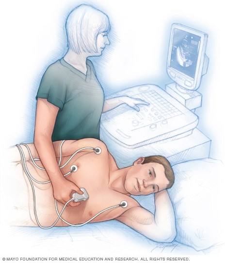
Transthoracic echocardiogram
In this standard type of echocardiogram:
- A technician (sonographer) spreads gel on a device (transducer).
- The sonographer presses the transducer firmly against your skin, aiming an ultrasound beam through your chest to your heart.
- The transducer records the sound wave echoes from your heart.
- A computer converts the echoes into moving images on a monitor.
If your lungs or ribs block the view, you may need a small amount of an enhancing agent injected through an intravenous (IV) line. The enhancing agent, which is generally safe and well tolerated, will make your heart's structures show up more clearly on a monitor.
Transesophageal echocardiogram
If your doctor wants more-detailed images or it's difficult to get a clear picture of your heart with a standard echocardiogram, your doctor may recommend a transesophageal echocardiogram.
In this procedure:
- Your throat will be numbed, and you'll be given medications to help you relax.
- A flexible tube containing a transducer is guided down your throat and into the tube connecting your mouth to your stomach (esophagus).
- The transducer records the sound wave echoes from your heart.
- A computer converts the echoes into detailed moving images of your heart, which your doctor can view on a monitor.
Doppler echocardiogram
Sound waves change pitch when they bounce off blood cells moving through your heart and blood vessels. These changes (Doppler signals) can help your doctor measure the speed and direction of the blood flow in your heart.
Doppler techniques are generally used in transthoracic and transesophageal echocardiograms. Doppler techniques can also be used to check blood flow problems and blood pressure in the arteries of your heart — which traditional ultrasound might not detect.
The blood flow shown on the monitor is colorized to help your doctor pinpoint any problems.